Ridpath recently retired from the ruminant diseases and immunology research unit, NADC/ARS/USDA, Ames, Iowa. Miller is a senior professional services veterinarian-dairy, Boehringer Ingelheim Vetmedica Inc., St. Joseph, Mo.
Cattle that have suffered from BVD infections are left with damaged immune systems. These wounded immune systems are subsequently less able to fight off infections initiated by other invading pathogens. This results in more infectious diseases and a drop in production, and this is where BVD inflicts major economic damage.
Hiding in the shadows
Infection with the viruses that cause BVD can result in either acute or persistent infections. Acute infections can occur in cattle starting from the second trimester in utero until the end of the animal’s lifespan. During acute infection, circulating white blood cells and immune tissues such as lymph nodes, thymus, and spleen are attacked and white blood cell counts are reduced.
Depending on the viral strain, the health status of the animal, and the presence of other pathogens, this depletion of the immune system may be accompanied by fever, diarrhea, or pneumonia, and some animals may succumb to disease. Those that are capable clear the virus within two weeks and clinical signs resolve.
Persistent infections occur when fetuses are exposed during the first and early second trimesters of gestation, before the immune system is fully functional. The subsequent damage to the developing immune system results in immunotolerance to the virus. This means that the immune system’s ability to recognize the infecting virus is destroyed. The immune system “tolerates” the virus and doesn’t eliminate it.
Persistently infected (PI) animals may appear stunted or completely normal and shed virus into the environment their entire lives. These PI animals are the source of most BVD outbreaks. The goal of testing programs is to detect and remove PI cattle.
Testing is most effective when it is part of an overall biosecurity strategy. The tests used for PI surveillance differ by the targets that they are detecting. Virus isolation (VI) focuses on replicating virus that can be detected by growth in cultured cells. Immunohistochemistry (IHC) and antigen capture ELISA (ACE) detect viral proteins. Polymerase chain reaction (PCR) tests detect the viral genome.
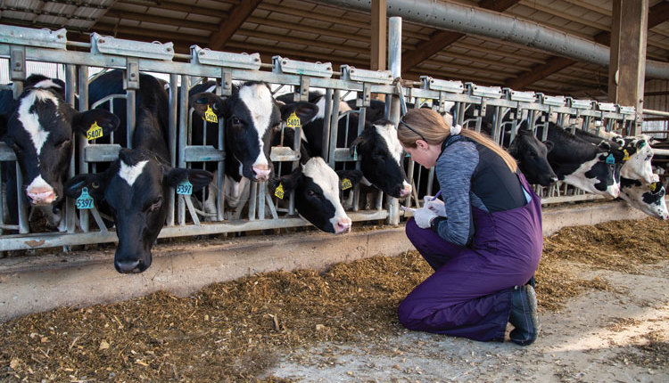
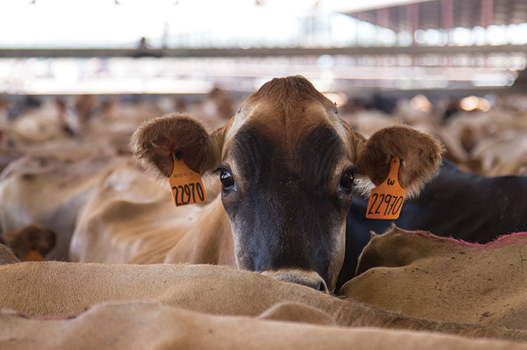
Because a PI animal has virus throughout their body, just about any tissue or bodily fluid can be used for testing. The most commonly used sources are blood and ear notches. However, IHC, which is dependent upon detection of virus in fixed and sectioned tissues on slides, cannot be used with blood samples.
Virus isolation is the most expensive and most time consuming testing option. VI is used primarily in cases with unusual presentations where a number of different viruses could be the causative agent. Following infection of cultured cells, virus specific antibodies or sequencing of the viral genomes present are used to identify the virus.
VI is useful in a situation in which PI animals are being born in multiple calf crops. VI followed by genotyping lets the producer know if the same virus is causing the problem or if multiple viruses are involved. VI is rarely used for routine surveillance.
The methods of choice for PI surveillance are IHC, ACE, and PCR. Unlike IHC, ACE and PCR samples may be pooled. While pooling of multiple samples is offered by many laboratories and lower costs of testing initially, pooling elevates the risk of missing PI animals. The larger the number of samples in a pool, the greater the risk of missing a PI animal.
While surveillance tests may be licensed by the United States Department of Agriculture (USDA), licensure is based on test results provided by the company that markets the test. The USDA does not have standardized testing criteria or test samples used to grant licenses, and there is no license available from the USDA for the laboratories that perform the tests.
One additional diagnostic test that may be done in the course of an outbreak is called virus neutralization (VN). VN tests detect antibodies in serum that have risen due to exposure to a virus. VI, IHC, ACE, and PCR are designed to detect current infections. VN tests are designed to detect prior infections.
Implement a control strategy
There are two different standards for licensing vaccines. One is the prevention of clinical disease following acute infections, and the other is the prevention of the birth of PI cattle. The level of protection needed to prevent PIs is much higher than that needed to prevent clinical disease following acute infection.
Two types of vaccine are available: modified live virus (MLV), which contains weakened or low virulence strains of virus; and killed virus (KV) in which all viral components have been deactivated. Both MLV and KV vaccines are effective. The KV vaccines are thought to be safer, and the MLV vaccines are thought to provide a longer lasting and broader immune response.
Vaccination is a powerful tool. However, it is not a silver bullet and cannot in and of itself provide complete protection against BVD. There are a number of reasons that vaccines fail (see sidebar), but even under ideal circumstances vaccination must be combined with testing and biosecurity for optimal BVD control.
First issue: Many faces of BVD
Last issue: Don’t bring BVD home with you
This issue: Strengthen your BVD fight
Why do BVD vaccines fail?
- A PI animal(s) is/are present in a herd.
- Insufficient time following vaccination for immune response to occur before exposure to virus (need at least two weeks).
- Animals were incubating disease at the time of vaccination.
- Interference when multiple vaccines are given concurrently.
- Maternal antibody interference in young calves.
- Failure to follow vaccine label directions.
• Failure to keep vaccines cold and in the dark.
• Use of soaps, detergents, disinfectants to clean the inside of multi-dose syringes used to administer modified-live vaccines (inactivates the vaccine).
• Failure to follow the labeled route administration or labeled dosage. - Use of expired vaccines.
- Failure to mix the vaccine properly and use a modified-live vaccine within 1 hour of mixing.
- Failure to follow up with a second dose of vaccine three to four weeks after initial dose when administering killed vaccines.
- Stress at the time of vaccine administration.
- Inadequate nutrition.
- Failure to protect vaccines from sunlight (heat) and extreme cold (freezing).
- Poor match between field strain and vaccine strain.