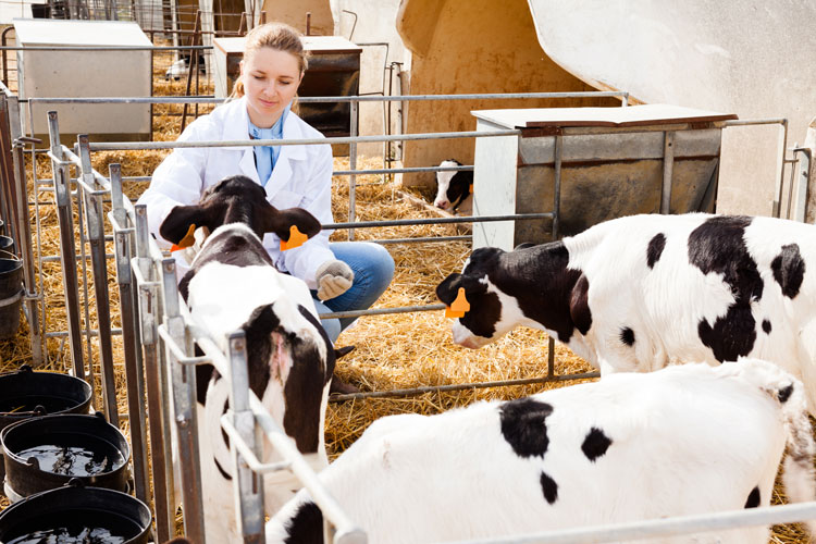
The author is a partner and large animal veterinarian at Thumb Veterinary Services in Deckerville, Mich.

We’ve probably all quipped the phrase, “If you have livestock, you’ll have dead stock, too.” It’s so true.

In many instances, we have a good notion as to the cause of death, but other times, we are at a loss for the why. History and symptoms generally narrow down our list of possibilities. Examples might include the Johne’s cow that has lost excessive body condition and developed chronic “pancake batter” loose manure for several weeks. She still eats fairly well, but the udder fill is diminishing. Many have also seen the typical listeriosis infected animal, with a history of progressive neurological signs, facial paralysis often on one side of the head, and circling in the pen, eventually becoming unable to rise.
I was taught 40 years ago that “common things happen commonly,” which is valid . . . but what about the uncommon?
It is tough on the dairy farm to always be expected to accurately diagnose disease. Should we record the death in the fresh cow pen as digestive? Metabolic? Injury? It is not always so easy.
Performing a necropsy on the farm allows a “look inside,” which, in many instances, is of great help recognizing and identifying the cause(s) of death. I recently participated in a necropsy master class offered by Zoetis along with fellow Michigan bovine practitioners. It was wonderful to join in the learning presented by seasoned bovine pathologists. I will share some of the take-home messages as we stood around in a ring and took a look inside several euthanized cows. Interesting cases were revealed from the inside!
Accurate and rapid diagnoses
Farmers today need fast disease detection. From newborn calves to adult cows, during an outbreak of disease, time matters! Early answers provide the path for treatment, prevention, and timely control. Accurate diagnoses are critical.
At the training, the seasoned faculty demonstrated to us the importance of being thorough by conducting a detailed, methodical exam from “lips to tail.” During herd outbreaks with infectious disease, selecting animals to analyze early in the course of illness, that are preferably untreated with antimicrobials, allows enhanced accurate pathogen detection. The timing of a necropsy is critical, as postmortem changes become apparent rapidly, especially in warm weather.
Our practice has always appreciated the value of timely postmortems. Actually, it is wild to recount all the memorable necropsy cases we have seen!
Allows for further study
At times, the farm necropsy does not provide all of the evidence needed for diagnoses. Examples may include the scouring newborn calf, which often exhibits few visual postmortem findings. Typically, dehydration causes the morbidity and mortality, but further histological changes can answer the why and what agent(s) were involved. Tissue submission at the time of necropsy can also be helpful in monitoring nutritional status or detection of toxins. Perhaps there is additional value as we advance our nutritional knowledge in disease resistance.
Tissue disease detection, or histological change, is considered the “gold standard” in pathology. We have become rather sophisticated over the last decade in detection of microbes, the viral and bacterial agents that can give trouble to our herds. Examples of live animal diagnostics might include polymerase chain reactions (PCRs), viral isolation, and bacteriology from milk, the gut, and the airways.
Often times, the “bugs” identified may or may not be causing the disease we witness in the herd. Some are considered normal flora with disease potential. Examples of this would include isolation of Rotavirus, Cryptosporidium, and Clostridial spp. in both healthy and sick young stock. Tissues submitted from necropsy can offer histological evidence of suspected disease.
The secret lies in the middle
Field necropsies provide a great opportunity for accurate detection of disease. Many deaths are more obvious and do not need further investigation, but timely postmortem exams provide learning opportunities for everyone caring for the herd. We all miss more from not looking than not knowing! Take the time to observe and ponder the whys. It matters.
Thank you to the Michigan Zoetis team for providing such valuable and interesting veterinary continuing education. Lifelong learning from great colleagues and super-sharp dairy farmers is so enjoyable!
I am also thankful for the much-needed precipitation scattered over our dairy farms. Hopefully timely rains fall when you need them in your part of the world. Take in the dog days of summer, relax, and enjoy the dairy life!


